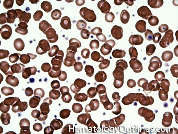
Thick area of a blood smear may give rise to overlapping RBCs (Pseudo-Rouleaux)
› Differential Diagnoses:
Plasma cell Neoplasms (e.g. plasma cell myeloma) Chronic liver disease with hypergammaglobulinemia Chronic infections Chronic inflammation Evaluating the wrong area of a slide (e.g. thick area of a blood smear with pseudo-rouleaux)
› Classic Immunophenotype:
N/A
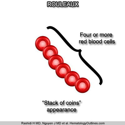
› Cartoon Image:
› Misc:
This finding must be evaluated in the appropriate section of the peripheral blood smear (where RBCs are usually not touching eachother)
Mostly due to reduced zeta potential (Note: The zeta potential between RBCs keeps them apart. Hence anything, usually relatively positively charged molecules such as immunoglobulins, that reduces this zeta potential will facilitate RBCs to aggregate or stack on top of each other)