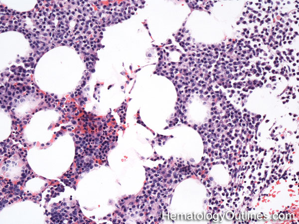
Usually presents as sheets or clusters of plasma cells in the bone marrow
Each plasma cell is 2-3x larger than a mature RBC
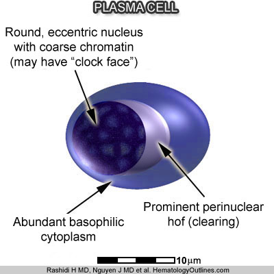
Round eccentrically placed nucleus with coarse chromatin (Nucleus may show a clock-faced chromatin pattern)
Abundant basophilic (blue) cytoplasm with prominent perinuclear hof (clearing)
Nucleoli are usually absent (some may have immature features such as prominent nucleoli. Other abnormal features may include nuclear pseudoinclusions known as "Dutcher Bodies")
› Normal % blood-PB, marrow-BM, lymphoid tissue-LN:
Plasma cell myeloma Monoclonal gammopathy of undetermined significance (MGUS) Lymphoplasmacytic Lymphoma Marginal zone lymphoma Plasmablastic lymphoma Chronic infection Autoimmune disorder Drug reaction
› Classic Immunophenotype:
CD138+
CD38 bright+
CD19-
CD56+ (common)
CD20-
Cytoplasmic light chain restricted (kappa only or lambda only)
› Cartoon Image:
Click and drag for direct comparison
› Misc:
The plasma cells in plasma cell myeloma (multiple myeloma) are monotypic (clonal) and typically involve the bone marrow as sheets or clusters of plasma cells. Monotypic refers to them being either kappa or lambda light chain restricted (as opposed to normal polytypic plasma cells which have a mix of kappa and lambda expressing plasma cells)
In Symptomatic Plasma Cell Myeloma, patients present with one or more of the "CRAB" findings: "C" for hyperCalcemia, "R" for renal insufficiency secondary to the disease, "A" for Anemia associated with the disease, and "B" for Bone Lesions (lytic)


