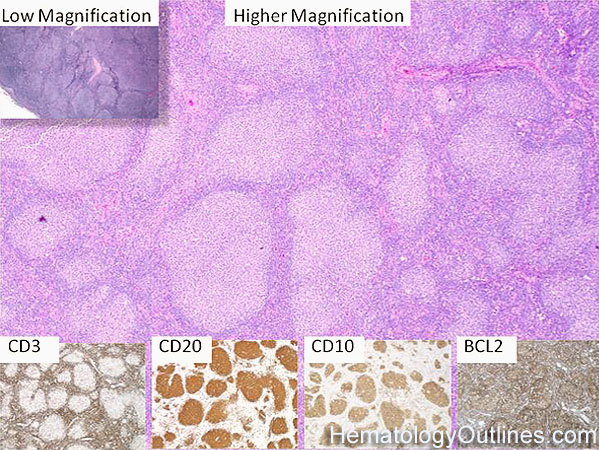
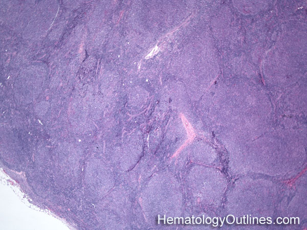
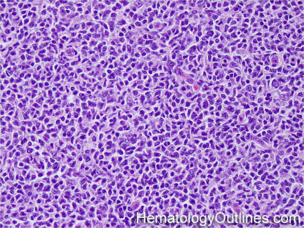
The lymph node is usually effaced (closed subcapsular sinus and loss of follicle architecture)
The lymphoid tissue is replaced by a back-to-back nodular (follicular) pattern of lymphoid proliferation with a diminished mantle zone
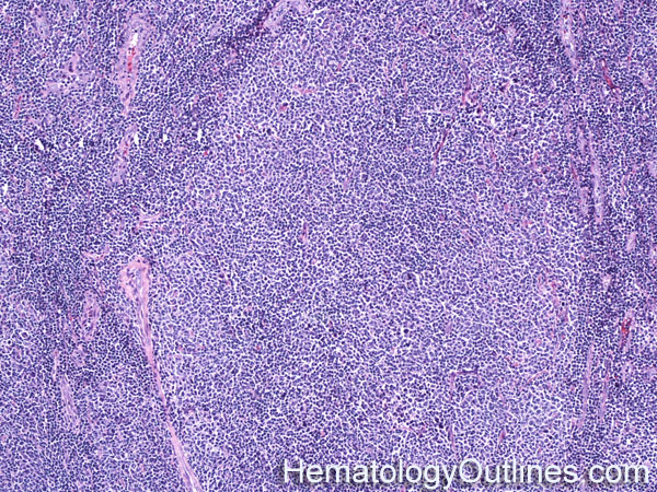
The majority of the neoplastic lymphocytes are located in the nodular areas and comprised of small cleaved (centrocytes) lymphocytes with rare scattered larger lymphocytes with multiple peripheralized nucleoli (centroblasts)
› Normal % blood-PB, marrow-BM, lymphoid tissue-LN:
Non-neoplastic reactive Lymph node Small Lymphocytic Lymphoma (SLL: The CLL equivalent in tissue) Mantle Cell Lymphoma Burkitt Lymphoma Diffuse Large B-cell Lymphoma
› Classic Immunophenotype:
CD19+
CD20+
CD10+
BCL6+
BCL2+
CD5-
› Cartoon Image:
› Misc:
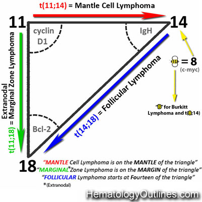
Associated with t(14;18) IgH:BCL2 genes
As opposed to Burkitt lymphoma which is also CD10+ with a Ki67 of ~100, the Ki67 (proliferative index) in most Follicular lymphomas are much lower.
Additionally, similar to other low grade lymphomas, the BCL2 protein (anti-apoptotic protein and not the gene) is typically overexpressed in most Follicular lymphomas. While, high grade lymphomas with increased apoptotic cells such as Burkitt lymphoma usually lack expression of the BCL2 protein.


