HematologyOutlines - Atlas
Primary Hematopoietic and Lymphoid Tissue ›› Bone Marrow ›› Abnormal
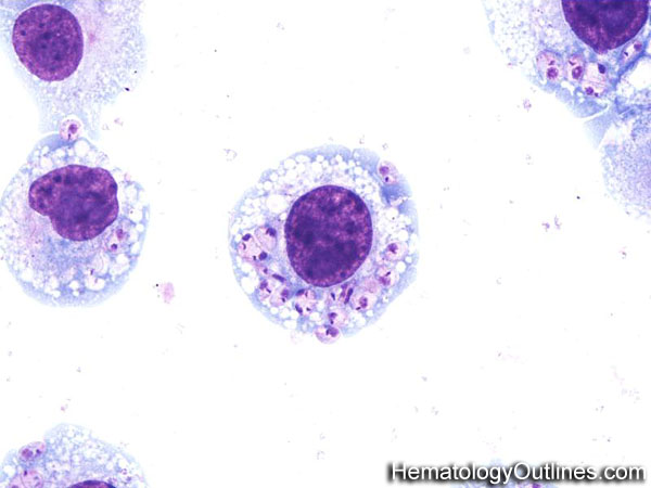
Leishmania in Histiocytes*
 Click Here for Full Size Click Here for Full Size
 › Microscopic Features:- Small round/oval parasitic forms with kinetoplasts are noted here within the cytoplasm of this histiocyte
 › Normal % blood-PB, marrow-BM, lymphoid tissue-LN:- PB: None
- BM: None
- LN: None
 › May Resemble:- Histoplasma in histiocytes
- Cellular debris in Histiocytes
- Bacteria in Histiocytes
 › Differential Diagnoses:
Leishmaniasis
Histoplasmosis
Cellular debris within histiocytes
Bacterial infection |
 › Classic Immunophenotype:
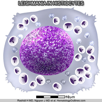
 › Cartoon Image:
 Click and drag

for direct comparison › Misc:- Histoplasma fungal forms lack kinetoplasts (small dark blue rod shaped structures within the organism) which are present in leishmania
|
|