HematologyOutlines - Atlas
Primary Hematopoietic and Lymphoid Tissue ›› Bone Marrow ›› Normal

Basophil (Mature)*


Click Here for Full Size


Microscopic Features:
  • 2-3x larger than a mature RBC
  • Low nuclear to cytoplasmic ratio (more cytoplasm than nucleus)
  • Nucleus is mature and usually with 2-3 lobes connected by thin chromatin filament
  • Nucleoli are absent
  • More cytoplasm with only secondary granules
  • Secondary (specific) granules are Basophilic (blue-violet)
  • The granules obscure the underlying nucleus
Normal % blood-PB, marrow-BM, lymphoid tissue-LN:
  • PB: Very Rare (5th most common WBC in blood, after neutrophils, lymphocytes, monocytes, and Eosinophils)
  • BM: Rare scattered
  • LN: None
May Resemble:
  • Eosinophil
  • Segmented Neutrophil
  • Pseudo-Pelger Huet Neutrophil
  • Band Neutrophil
  • Neutrophil with toxic granules
  • Promyelocyte
  • Mast Cell
Differential Diagnoses:

Increased in:
CML accelerated phase
AML (rare variants)
Infection (e.g. Varicella)
Hypersensitivity reactions

Classic Immunophenotype:
  • CD45dim+
  • Low SSC (Side light scatter)
  • CD11b+
  • CD13+
  • CD16-
  • CD15-
Cartoon Image:


Click and drag
RBC
for direct comparison

Misc:
  • Increased basophils may be associated with some myeloid neoplasms such as CML, some infections or some hypersensitivity reactions Basophils and Mast cells are distinct from one another but share some similar morphologic and functional aspects. As opposed to basophils which can be seen in the peripheral blood, mast cells are only seen in tissue and are absent from the peripheral blood. 
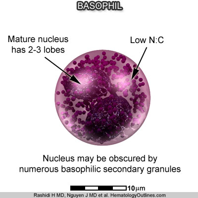
Additionally, the nucleus of the mast cell is usually round and not segemented as opposed to the basophils' segmented (usually bi-lobed) nucleus. The granules of basophils are more heterogenous and overlap the nucleus while the granules of mast cells are more uniform and less often cover the nucleus. (For more details, please see the mast cell entry under the Glossary section.)