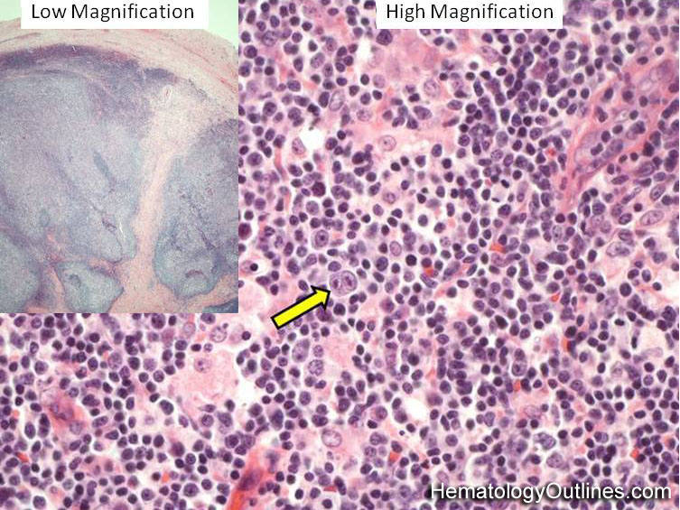
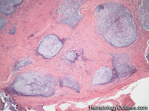
The lymph node is usually effaced (closed subcapsular sinus and loss of normal follicle architecture)
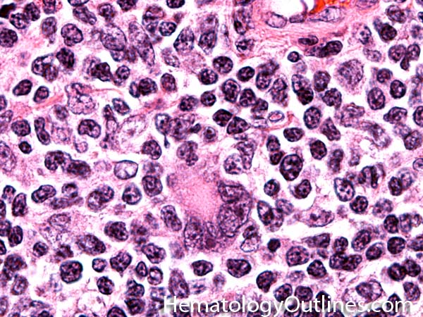
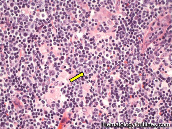
Background is usually Lymphohistiocytic (mix of small lymphocytes and larger histiocytes with abundant pink cytoplasm)
Scattered background neoplastic cells are the Reed-Sternberg (RS) cells (2 or more nuclei with very large nucleoli) and Hodgkin cells (single large nucleus with large nucleolus)
› Normal % blood-PB, marrow-BM, lymphoid tissue-LN:
PB: N/A
BM: N/A
LN: N/A
› May Resemble:
Non-neoplastic reactive Lymph node
Follicular Lymphoma
Diffuse Large B-cell Lymphoma (esp. the T-cell/Histiocyte Rich Large B-cell Lymphoma subtype)
› Differential Diagnoses:
Diffuse Large B-cell Lymphoma (esp. The T-cell/Histiocyte Rich Large B-cell Lymphoma subtype) Non-neoplastic reactive Lymph node Follicular Lymphoma
› Classic Immunophenotype:
RS cells: CD30+, CD15+/-, PAX5 dim+ (a B-cell marker), and most are OCT2, BOB1 and CD20 negative
Also, as opposed to most hematopoietic cells which are CD45+, RS cells are CD45 negative (CD45 is also known as LCA or Leukocyte Common Antigen)
› Misc:
Hodgkin usually presents in supra diaphragmatic lymph nodes or lymphoid areas
Usually thought to be EBV associated
Classical Hodgkin Lymphoma has 4 subtypes: Nodular Sclerosing, Mixed Cellularity, Lymphocyte-Rich, and Lymphocyte depleted.