If increased in blood or increased numbers in bone marrow: Chronic Myelomonocytic Leukemia Autoimmune disorders (sometimes) Chronic infections (e.g. CMV, tuberculosis, etc.)
› Classic Immunophenotype:
CD45+
Intermediate SSC (Side light scatter)
CD14+
CD4dim+
CD64+
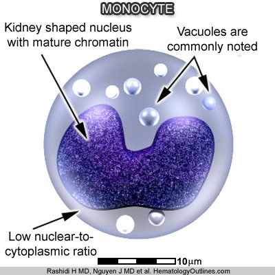
› Cartoon Image:
Click and drag for direct comparison
› Misc:
Monocytes leave the blood and enter tissue to become macrophages/histiocytes


