Primary Hematopoietic and Lymphoid Tissue ›› Bone Marrow ›› Normal

Megakaryocyte*


Click Here for Full Size


Microscopic Features:
  • 10-30x larger than a mature RBC (Largest hematopoeitic cell)
  • Lower nuclear to cytoplasmic ratio because of increased cytoplasm
  • Multilobated Nucleus (2-8 lobes on top of each other) with mature chromatin (clumped)
  • Nucleoli are absent
  • Cytoplasm is abundant and light blue and usually lacks granules
Normal % blood-PB, marrow-BM, lymphoid tissue-LN:
  • PB: None
  • BM: Scattered
  • LN: None
May Resemble:
  • Osteoclast
  • Histiocytes and macrophages (specially multinucleated forms)
  • Hodgkin Lymphoma Reed Sternberg cell [Compare]
  • Metastatic tumor cells
Differential Diagnoses:

Increased numbers in bone marrow:
Peripheral destruction of platelets (e.g. ITP) may lead to megakaryocytic hyperplasia in the bone marrow
Myeloproliferative neoplasms
Myelodysplastic Syndrome (specifically MDS with isolated 5q deletion)

Classic Immunophenotype:
  • CD41+
  • CD42+
  • CD61+
Cartoon Image:


Click and drag
RBC
for direct comparison

Misc:
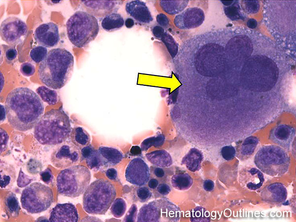
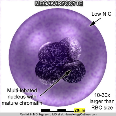
  • Megakaryocytes give rise to the platelets which circulate in blood. Megakaryocytes are not present in the peripheral blood. Under normal circumstances they reside in the bone marrow and when the marrow is unable to produce (e.g. Primary Myelofibrosis), they may be seen in other organs such as the spleen as part of Extramedullary Hematopoeisis


Content Editors/Website Administrators:
Hooman H. Rashidi, MD; John C. Nguyen, MD