Primary Hematopoietic and Lymphoid Tissue ›› Bone Marrow ›› Myeloid ›› Megakaryocytes & Precursors ›› Abnormal
Hypolobated Megakaryocytes (Increased)
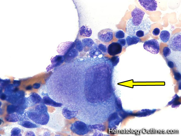

 ›Microscopic Features:
- 8-10x larger than a mature RBC
- Lower nuclear to cytoplasmic ratio because of increased cytoplasm
- Many are hypolobated (1-2 lobes) with a mature (clumped chromatin) Nucleus
- Nucleoli are absent
- Cytoplasm is abundant and light blue and usually lacks granules
 ›Normal % blood-PB, marrow-BM, lymphoid tissue-LN:
- PB: None
- BM: Rare Scattered
- LN: None
 ›May Resemble:
- Megakaryocyte (normal)
- Osteoclast
- Histiocytes and macrophages (specially multinucleated forms)
- Osteoblast
- Hodgkin Lymphoma Reed Sternberg cell
- Metastatic tumor cells
- Myelocyte
 ›Differential Diagnoses:
Increased numbers in bone marrow:
MDS
Reactive increase secondary to peripheral destruction of platelets
Post Chemotherapy
Toxin-induced
MDS
Reactive increase secondary to peripheral destruction of platelets
Post Chemotherapy
Toxin-induced
 ›Classic Immunophenotype:
- CD41+
- CD42+
- CD61+
 ›Cartoon Image:
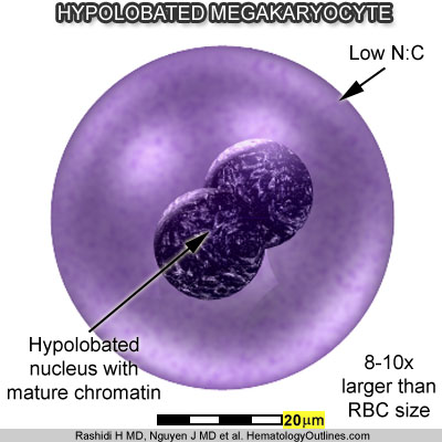
Click and drag for direct comparison
for direct comparison ›Misc:
Hypolobated Megakaryocytes may be dysplastic and associated with a myelodysplastic syndrome (MDS) or newly forming megakaryocytes that are the result of a peripheral destruction of platelets (e.g. ITP)