Secondary Lymphoid Tissues ›› Lymph Node ›› Neoplastic: Hodgkin Lymphoma
Classical Hodgkin Lymphoma
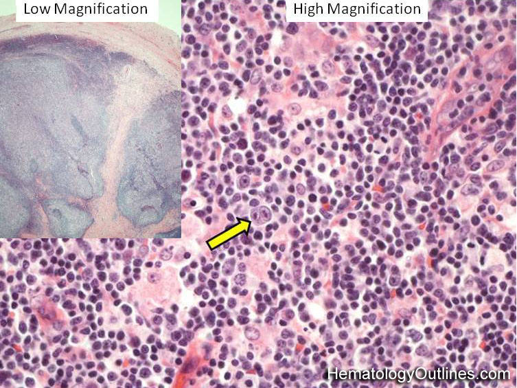

 ›Microscopic Features:
- The lymph node is usually effaced (closed subcapsular sinus and loss of normal follicle architecture)
- Background is usually Lymphohistiocytic (mix of small lymphocytes and larger histiocytes with abundant pink cytoplasm)
- Scattered background neoplastic cells are the Reed-Sternberg (RS) cells (2 or more nuclei with very large nucleoli) and Hodgkin cells (single large nucleus with large nucleolus)
 ›Normal % blood-PB, marrow-BM, lymphoid tissue-LN:
- PB: N/A
- BM: N/A
- LN: N/A
 ›May Resemble:
- Non-neoplastic reactive Lymph node
- Follicular Lymphoma
- Diffuse Large B-cell Lymphoma (esp. the T-cell/Histiocyte Rich Large B-cell Lymphoma subtype)
 ›Differential Diagnoses:
Diffuse Large B-cell Lymphoma (esp. The T-cell/Histiocyte Rich Large B-cell Lymphoma subtype)
Non-neoplastic reactive Lymph node
Follicular Lymphoma
Non-neoplastic reactive Lymph node
Follicular Lymphoma
 ›Classic Immunophenotype:
- RS cells: CD30+, CD15+/-, PAX5 dim+ (a B-cell marker), and most are OCT2, BOB1 and CD20 negative
- Also, as opposed to most hematopoietic cells which are CD45+, RS cells are CD45 negative (CD45 is also known as LCA or Leukocyte Common Antigen)
 ›Misc:
Hodgkin usually presents in supra diaphragmatic lymph nodes or lymphoid areas
Usually thought to be EBV associated
Classical Hodgkin Lymphoma has 4 subtypes: Nodular Sclerosing, Mixed Cellularity, Lymphocyte-Rich, and Lymphocyte depleted.
Usually thought to be EBV associated
Classical Hodgkin Lymphoma has 4 subtypes: Nodular Sclerosing, Mixed Cellularity, Lymphocyte-Rich, and Lymphocyte depleted.