Primary Hematopoietic and Lymphoid Tissue ›› Bone Marrow ›› Myeloid ›› Erythroid (RBCs & Precursors) ›› Normal
Polychromatophilic RBC
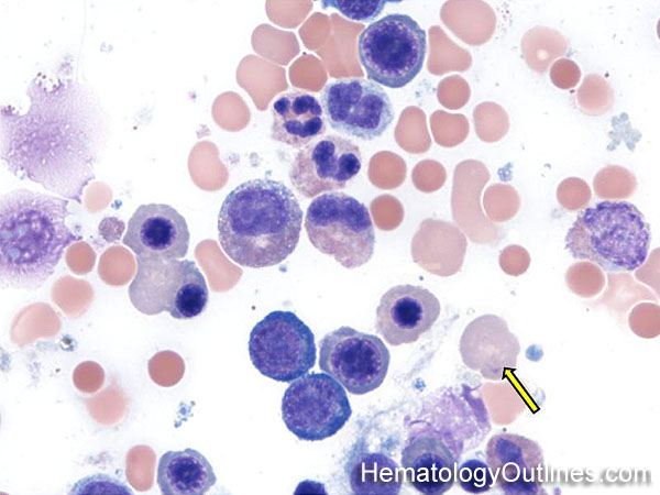

 ›Microscopic Features:
- Slightly larger than normal mature RBCs
- Lacks a nucleus
- Cytoplasm is grayish-orange in color
- It's called a Reticulocyte when the precipitated RNA can be highlighted with supravital stain
 ›Normal % blood-PB, marrow-BM, lymphoid tissue-LN:
- PB: Rare Scattered
- BM: Scattered
- LN: None
 ›May Resemble:
- Polychromatophilic RBC (Wright stain) may resemble:
- Mature RBC
- Macrocytic RBC
- --------------------------------------
- Reticulocyte (supravital stain) may resemble:
- Heinz bodies
- RBC with Stain artifact
 ›Differential Diagnoses:
Increased in:
Newborns
In response to certain anemias (e.g. hemolytic anemia)
In response to acute blood loss
In response to a recovering marrow (post therapy)
Reactive Erythroid hyperplasia
Post-erythropoietin therapy
Polycythemia Vera
Newborns
In response to certain anemias (e.g. hemolytic anemia)
In response to acute blood loss
In response to a recovering marrow (post therapy)
Reactive Erythroid hyperplasia
Post-erythropoietin therapy
Polycythemia Vera
 ›Classic Immunophenotype:
- CD45-
- CD117-
- CD235a (Glycophorin A) +
- CD71-
 ›Cartoon Image:
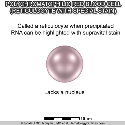
Click and drag for direct comparison
for direct comparison ›Misc:
They lack a nucleus. Scattered cells can also be seen in the blood. The grayish color of the cytoplasm is due to its increase number of RNA moelcules since these cells are still less mature than the fully mature RBC
Note: As Erythroid precursors mature, the color of their cytoplasm changes from blue to gray to red-orange (this is due to the increase in the number of hemoglobin molecules within the red cells).
Note: As Erythroid precursors mature, the color of their cytoplasm changes from blue to gray to red-orange (this is due to the increase in the number of hemoglobin molecules within the red cells).