Secondary Lymphoid Tissues ›› Spleen ›› Normal
Normal Spleen
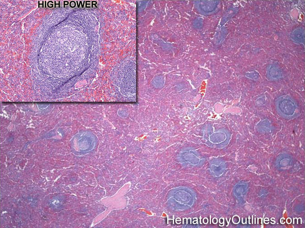

 ›Microscopic Features:
- Spleen is divided into 2 main zones: Red pulp and white pulp
- The white pulp contains the lymphoid cells and appears as small blue/purple aggregates of lymphoid tissue
- The red pulp contains the sinusoids along with scattered hematopoietic cells and appears red in the image above
 ›Normal % blood-PB, marrow-BM, lymphoid tissue-LN:
- PB: N/A
- BM: N/A
- LN: N/A
 ›May Resemble:
 ›Differential Diagnoses:
Reactive lymphoid hyperplasia
Infection
Splenic Marginal Zone Lymphoma
Hairy Cell Leukemia
Follicular Lymphoma
Infection
Splenic Marginal Zone Lymphoma
Hairy Cell Leukemia
Follicular Lymphoma
 ›Classic Immunophenotype:
- "T"-cells have the "Tiny" CDs: CD2, CD3, CD4, CD5, CD7, CD8
- "B"-cells have the "Bigger" CDs: CD19, CD20, CD22
 ›Misc:
In Splenic Marginal Zone Lymphoma, the neoplastic cells involve the white pulp area of the Spleen (as opposed to Hairy Cell Leukemia which involves the red pulp area)