Primary Hematopoietic and Lymphoid Tissue ›› Bone Marrow ›› Myeloid ›› Monocytes & Precursors ›› Normal
Monocyte
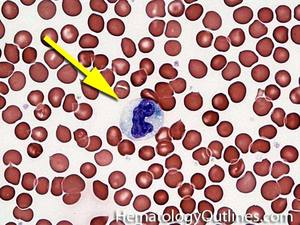

 ›Microscopic Features:
- 3-4x larger than a mature RBC
- Lower nuclear to cytoplasmic ratio because of increased cytoplasm
- Kidney shaped Indented Nucleus with mature chromatin (clumped)
- Nucleoli are absent
- Cytoplasm is abundant, gray to pale blue and with rare to no granules
- vacuoles are commonly noted in cytoplasm
 ›Normal % blood-PB, marrow-BM, lymphoid tissue-LN:
- PB: Monocytes are the 3rd most common WBC in the blood (after neutrophils and lymphocytes)
- BM: Scattered monocytes can be seen in the bone marrow (some in the form of histiocytes/macrophages)
- LN: Scattered monocytes can be seen in the bone marrow (some in the form of histiocytes/macrophages)
 ›May Resemble:
 ›Differential Diagnoses:
If increased in blood or increased numbers in bone marrow:
Chronic Myelomonocytic Leukemia
Autoimmune disorders (sometimes)
Chronic infections (e.g. CMV, tuberculosis, etc.)
Chronic Myelomonocytic Leukemia
Autoimmune disorders (sometimes)
Chronic infections (e.g. CMV, tuberculosis, etc.)
 ›Classic Immunophenotype:
- CD45+
- Intermediate SSC (Side light scatter)
- CD14+
- CD4dim+
- CD64+
 ›Cartoon Image:
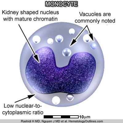
Click and drag for direct comparison
for direct comparison ›Misc:
Monocytes leave the blood and enter tissue to become macrophages/histiocytes