Primary Hematopoietic and Lymphoid Tissue ›› Bone Marrow ›› Lymphoid ›› T-cells ›› Abnormal
Large Granular Lymphocyte (LGL)
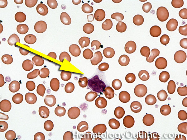

 ›Microscopic Features:
- 2-3x larger than a mature RBC
- Low nuclear to cytoplasmic ratio but with
- Round mature Nucleus (clumped chromatin)
- Nucleoli are absent
- Cytoplasm is abundant, light blue
- Pink/red granules in the cytoplasm
 ›Normal % blood-PB, marrow-BM, lymphoid tissue-LN:
- PB: Rare to None
- BM: Rare to None
- LN: None
 ›May Resemble:
 ›Differential Diagnoses:
If increased in blood or bone marrow:
Infection
Neoplastic (specifically LGL leukemia)
Drug-induced
Autoimmune disorders
Infection
Neoplastic (specifically LGL leukemia)
Drug-induced
Autoimmune disorders
 ›Classic Immunophenotype:
- CD2+
- CD3+
- CD8+
- CD5+
- CD7+
- CD56+
 ›Cartoon Image:

Click and drag for direct comparison
for direct comparison ›Misc:
Must rule out reactive conditions (e.g. infection, etc.) before considering LGL leukemia as a diagnosis. LGL leukemias will also show a clonal T-cell population through TCR gene rearrangement studies