Secondary Lymphoid Tissues ›› Mucosa-Associated Lymphoid Tissue or MALT ›› MALT ›› Abnormal
MALT Lymphoma (e.g. Gastric)


 ›Microscopic Features:
- Small- to intermediate-sized lymphocytes (many with monocytoid features which show increased amount of cytoplasm)
- Lymphoepithelial lesions (neoplastic lymphocytes infiltrating and destroying the epithelial glandular tissue)
 ›Normal % blood-PB, marrow-BM, lymphoid tissue-LN:
- PB: None
- BM: None
- LN: None
 ›May Resemble:
- H. pylori gastritis
- Follicular Lymphoma
- Diffuse Large B-cell Lymphoma
- Lymphoplasmacytic lymphoma
- Small Lymphocytic Lymphoma
- Mantle Cell Lymphoma
 ›Differential Diagnoses:
Infection (specifically H. Pylori in stomach)
Mantle cell lymphoma
Follicular lymphoma
Lymphoplasmacytic lymphoma
Diffuse Large B-cell Lymphoma
Mantle cell lymphoma
Follicular lymphoma
Lymphoplasmacytic lymphoma
Diffuse Large B-cell Lymphoma
 ›Classic Immunophenotype:
- CD19+
- CD20+
- CD5-
- CD10-
- CD23-
- BCL1-
 ›Cartoon Image:
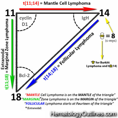
 ›Misc:
Gastric MALT lymphoma is usually associated with H. pylori
Note: Extranodal MALT lymphomas are more common in areas of the body that usually lack normal MALT lymphoid tissue (e.g.gastric, etc.)
Note: Extranodal MALT lymphomas are more common in areas of the body that usually lack normal MALT lymphoid tissue (e.g.gastric, etc.)